A REVIEW OF 10, 000 CASES TREATED BY MONOCLONAL ANTIBODIES
Dres. Gregorio Skromne Kadlubik y Ricardo Hidalgo R. Laboratorio de Radionúclidos
Departamento de Fisiología. Facultad de Medicina. UNAM. Ciudad de México, México
ABSTRACT:
10, 000 human cases of cysticercosis were treated with radionuclides in the last 14 years . An stadistical evaluation of localization of the cyst; number of them; main symtoms and signs; laboratory and gabinet findings; improvement and dissaparence of the illnes; certification by laboratory, gabinet and histophatological findings after treatment: socioeconomical consideration and an special consideration about ocular cysticercosis are revisited in this the most larger stadistical experience in the world about human cysticercosis.
MATERIAL AND METHODS:
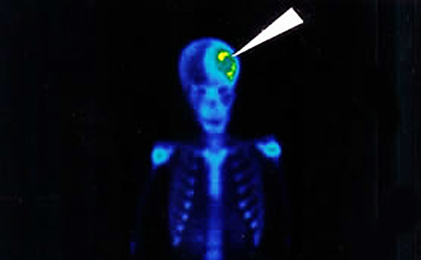
From March 1978 to March 1992 we studied a studied a total of 10, 000 patient with cysticercosis (most of then cerebral ) with an average of 16 (sixteen) patient a week. All the patient have a clinical evaluation, with inmunological test, cranial roentgenogram, electroencefalography, computed tomography scan and radioinmmunoscan prior and treatmen. The principles and method of radioinmmunoscan and radioinmmunotreatment were published elsewhere (1 , 2) .
The patient were seen a time from 6 months to 5 years follow-up. The results of this review are based in the stadistical revision of this material.
RESULTS AND DISCUTION:
9215 cases were of adults and anly 785 of children.
The minimal age was a child of only 17 days old (trans-placental infestation?) and the maximal age was a lady of 89 years.
The average age of clinical manifestation of the illnes were 37 years. The mayor number of patient were males (5930) versus 4, 070 of females. (Its probably have to be with higienical habits and home meats in the patients) . A few cases were from U.S.A , South-America (Colombia and Bolivia) , Centro-America (Nicaragua) & India, But basically the stadistic is based in Mexican patients (9969 cases) . The patient came from different institutions, or Physicians (9969 cases) . The patient came from different institutions, or Physicians.
 Table No. 1 present the casuistic of localization of cyst: most of them of course in brain, but we found cyst in themost curious localization (kidney, heart, etc.) and in several organs (brain and muscle, etc. ) . In brain multiple localization were the rule and the racemic from were the less present (see table No. 2) . In spinal cord medulla and cerebelum we find alse cyst in 5 cases (see table No. 3) .
The number of cyst is alse tabuled in table No. 3 The combination of cerebral localization are tabuled in table No. 4 in “X” and “Y” axis. The main symptoms and signs in cerebral cysticercosis are putted in table No. 5 . The most common symptoms find were headhache and the per cent of positivity with laboratory and gabinet studies in present in table No. 6 . False negative is the reciprocal of this cifres. In table No. 7 is present the per cent of improvement and dessaparence of the illness in the patients and in the last table (No. 8) in seen the laboratory, gabinet and histophatological findinge of the cysticercosis patient after radioinmmuno treatment.
Other remarkable points of the study are the socioeconomical consideration that we find and are the following:
1.-30% of the patients have reinfestations; maybe because the nonhigienical social conditions of their habitat persist .
2.- Social groups have different per cent of cysticercosis morbility . The mayor frecuency appers in soial medial level (60%) of the cases and not in low economical level (60%) of the cases and not in low economical level we expect (only 30%) maybe because they have more antibodies that the rest of population if the patient survive the first years of life; or maybe because the meat with more posibilities of infestation is not in their economical posibilities (strawberries, etc) of eating . The high society patient have only the 10% of the cases (logicall consideret the teoretical higienical level they live.).
3.-In our casuistic 40% of cyst are alive; and 60% are calcified .
4.- 13% of the patient have been previoualy treated with prazicuantal with many complications and with a poor clinical response that requiered a second treatment with radioinmmuno methods.
5.- 10% of the cases have previously surgery for cranial hipertension but a cyst blocket the valves coloqued in radioinmmunotreatment desing .
6.- 7% of the cases have been treated with an anticolinergic drug previously by other authors; causing hepatic damage and without improvement; that requiered a second treatment with radioimmuno method.
7.-Nobody of our patient have intolerance nor radiotoxicity with the radioimmunotreatment; and in all of them we try to have pure cases without contamination of anther drugs or treatments during is entire follow-up
8.- The avarage cost of the treatmen was $300.00 US Dlls. Per do se and don’t requiere hospitalization . Finally the consideration about the cases of ocular cysticercosis are the following:
1.- In right eye we fiund 3 cases .
2.- In left eye we find 5 cases .
3.- No bilateral cases we have seen till today .
4.- In every cases only ane cyst was seen in one eye.
5.- The cyst is radiolised and so is seen by oftalmoscophy study.
6.- All the cyst were radiolised with good results.
AKNOWLEDWMENT:
The following institutions and physicians referred patients for radioimmunotreatment that the authors to thank : Instituto de Seguridad y Servicios Sociales de los Trabajadores del Estado: Hospitales 20 de Noviembre, Fernando Quiroz, López Mateos, Darío Fernández, 1 de Octubre, Regional of San Luis Potosí and General of Toluca. Secretaría de Salubridad y Asistencia: Hospitales “Juárez”, Fray Bernadino Alvarez, and Instituto de Enfermedades Tropicales. Instituto Mexicano del Seguro Social: Hospital General Centro Médico La Raza and Puebla. Hospital Central de Petróleos Mexicanos, Hospital de Jesús. México, D.F., Centro Médico del Bajío, León, Gto.; Medical Center, Houston and Dr. César Celis González and Dr. Jesús Aguilar.
REFERENCES:
1.- Skromne-Kadlubik G.; Celis C.; Ferez A.: Cysticercosis of the nervous system : Diagnosis by means of specific radioimmuncs can .
Ann. Neurol . 1977, 2: 343.
2.- Skromne –Kadlubik G., Celis C. Cysticercosis of the Nervous system: treatment by means of Specific Internacional Radiation . Arch . of Neurology. 1981. 38: 288.
|
TABLE No. 1 |
|
|
CYST LOCALIZATION ON DIFERENT HUMANS ORGAN AND TISUES: |
|
|
BRAIN
|
9,782 |
|
CASES MUSCLES |
100* |
|
SUBCUTANEOUS TISUE |
90* |
|
KIDNEY |
2* |
|
HEART |
1* |
|
SUBMAXILARY SALIVAL GLANDS |
3* |
|
EYES |
8* |
|
LIVER |
1* |
|
TWO OR MORE ORGANS AND TISSUES |
13* |
|
Total: 10,000 cases |
|
|
*brain and muscle; brain and subcutancous tissue. |
|
|
Table No. 2 |
||||
|
CYSTICERCOSIS OF NERVOUS SYSTEM . LOCALIZATION: |
||||
|
LOCALIZATION |
RIGHT |
LEFT |
GLOBAL |
% |
|
FRONTAL LOBE |
392 |
587 |
979 |
10 |
|
PARIETAL LOBE |
41,122 |
2,741 |
6,853 |
70 |
|
OCCIPITAL LOBE |
489 |
488 |
977 |
10 |
|
TEMPORAL LOBE |
284 |
685 |
969 |
10 |
|
SUPRASELLAR |
2 |
|||
|
MEDULLA |
1* |
|||
|
SPINAL CORD |
3* |
|||
|
INTRATECAL |
10 |
|||
|
SUBTOTAL |
5,277 |
4,501 |
||
|
NOTES : * CONSIDERED OUTSIDE OF NERVOUS CENTRAL SYSTEM |
||||
|
+ EIGTH CASES HAVE ALSO EXTRA-CEREBRAL CYST SIMULTANSOUS |
||||
|
++ MULTIPLE LOCALIZATION WERE THE RULS IN CENTRAL NERVOUS SYS – TEM. ONLY 30% OF THE CASES WERE UNIQUE (SEE TABLE 3) |
||||
|
+++ THE MOST IMPORTANT CLINICAL AND REC (ELECTROENCEFALOGRAPHIC) – MANIFESTATION WERE CONSIDERED TO AGGREED WITH THE TOPOGRAPIC- LOCALIZATION OF THE CYST. FOR STADISTIC |
||||
|
++++ WHEN TWO OR MORE CYST WERE IN DIFFERENT LOBES WE STADISTICALLY |
||||
|
+++++ 20 % OF THE CASES WERE RACEMIC FROM OF CYSTICERCOSIS |
||||
|
Table No. 3 |
|
|
NUMBER OF CYSTICERCOS IN NERVOUS SYSTEM |
|
|
NUMBER OF CYST |
% OF TOTAL |
|
1 |
30 |
|
2 |
50* |
|
3-4 |
10** |
|
4 |
3*** |
|
5 a 10 |
5**** |
|
Más de 10 |
2**** |
|
Notes: |
|
|
(*) ALL THIS CASES WERE IN THE SAME LOBE WHEN TEEY APPERED EXAMPLE: TWO RIGHT FRONTAL OR TWO LEFT TEMPORAL |
|
|
(**) ALI THIS CASER WERE BILATERAL. THE MOST COMUN RIGHT PARIETAL/LEFT TEMPORAL |
|
|
(***) ALL THIS CASES WERE BILATERAL. THE MOST COMUN RIGHT PARIETAL/LEFT TEMPORAL |
|
|
(****) ALL THIS CASES WERE BILATERAL . ONE OF THEM HAVE ONE CYST IN CEREBELLUM |
|
|
Table No. 4 |
|||||
|
LOCALIZATION OF CYST IN CENTRAL NERVOUS SYSTEM |
|||||
|
FRONTAL |
TEMPORAL LEFT |
PARIETAL LEFT |
OCCIPITAL LEFT |
OTHER LOCALIZATION |
|
|
FRONTAL |
075 |
92 |
188 |
92 |
1 muscle |
|
TEMPORAL |
122 |
40 |
246 |
12 |
1 muscle |
|
PARIETAL |
92 |
322 |
78 |
33 |
1 subcutaneous |
|
OCCIPITAL |
47 |
17 |
31 |
119 |
1 muscle |
|
OTHER |
1 muscle |
1 cerebellum 1 muscle |
1 muscle |
1 muscle |
1 muscle |
|
Table No. 5 |
|
|
MAIN SYMTOMS AND SIGNS CEREBRAL CYSTICERCOSIS |
|
|
SYMTOMS |
% OF CASES |
|
HEADHACHE |
60 |
|
SEIZURES |
25 |
|
ENDOCRANSAL HYPERTENSION |
12 |
|
PSEUDOTUMOR |
1 |
|
MEDULAR SCHOK |
1 |
|
MISCELLANEOUS (Blind , coma, extrasistoles, etc) |
1 |
|
Table No. 6 |
|
|
LABORATORIY AND GABINET STUDIES IN CEREBRAL CYSTICERCOSIS |
|
|
Studies |
% OF POSITIVITY |
|
ELECTROENCHEFALOGRAPHY |
22 |
|
CRANEAL X RAY |
10* |
|
COMPUTARIZED AXIAL TOMOGRAPHY |
65 |
|
INMUNOLOGYCAL TEST |
96 |
|
RADIOINMUNOSCAN |
65 |
|
*Changes of endocraneal hypertención, clacifications, etc. |
|
|
Tabla No. 7 |
|
|
PORCENT OF IMPROVEMENT AND DISSAPARENCE OF SYMPTOMS AND SYNDROMS |
|
|
SYMPTOMS AND SYNDROMS |
% |
|
SEIZURES |
96 |
|
ENDOCRANIAL HIPERTENSION |
97 |
|
PSEUDO-TUMOR |
100 |
|
SCHOKC SPINAL |
25 |
|
HEADHACHE |
85 |
|
COMA |
50 |
|
BASAL ARACNOIDITIS |
50 |
|
OTHERS (Sub-cutansous, eyes, ect.) |
90 |
|
Table No. 8 |
|
|
LABORATORY, GABINET AND HISTOPHATOLOGICAL FINDINGS IN CYSTICERCOSIS AFTER RADIOINMUNOTRENTMENT |
|
|
STUDIES |
% OF DISSAPARENCE OF SIGNS: OR CYS |
|
ELECTROENCEPHALOGRAPHY |
50 |
|
X RAY |
60 |
|
COMPUTARAIZED AXIAL TOMOGRAPHY |
90 |
|
INMUNOLOGICAL TEST |
96 |
|
RADIOINMUNOSCA |
50 |
|
SUBCUTANEOUS BYOPSY |
100* |
|
NEUROSURGERY |
100** |
|
NERCROPSY |
100*** |
|
Notes: |
|
|
* 17 CASES ALL WITH RADIO LYSIS. |
|
|
** 6 CASES ALL WITH RADIO LYSIS. |
|
|
*** 2 CASES : ONE DIED BY TUBERCULOSIS. AND OTHER DIED BY DISENTERY. ( Cyst radiolysed) |
|


